These HR/NHEJ kits are cell based, meaning they have the HR or NHEJ GFP reporters pre-installed. A single plasmid (I-Sce1) is transfected to activate repair. Please see this table for a summary and comparison/application of each kit.
Transfection-based HR and NHEJ kits use transient transfections to make a pool of cells containing the DNA reporters to allow the investigator to interrogate the 2 major DNA repair pathways. The advantages are:
It uses GFP readout to measure the efficiency of repair.
There is zero background when repair is blocked or inhibited
The reporters for HR or NHEJ can be installed in virtually any cell line that can be transiently transfect
It is rapid and robust
It is ideal for drug screens to find novel inhibitors of HR/NHEJ
The kits are highly specific and can only report HR or NHEJ
Readout GFP can be done by FACS or Imaging
Entire pathway can be followed by live imaging in viable cells
Ideal to track repair kinetics at the single cell level
The Kit is designed to allow quick and specific detection of DNA gyrase. This kit facilitates the purification and characterization of type II topoisomerase enzymes (DNA Gyrase) and contains all reagents necessary for routine assays of type II enzymes that either have or do not have the ability to supercoil.
If you want to order this product, please contact us.
The Kit is designed to allow quick and specific detection of DNA Gyrase. This kit facilitates the purification and characterization of type II topoisomerase enzymes (DNA Gyrase) and contains all reagents necessary for routine assays of type II enzymes that either have or do not have the ability to supercoil.
Gyrase Assay Kit Contents
-Kinetoplast DNA [kDNA] sufficient for 100 assays
-Marker linear kDNA
-Marker decatenated kDNA (topo II treated)
-10X Gyrase assay buffer
-Gyrase stop and load buffer
–Detailed instruction manual
Application Notes for DNA Gyrase Assay Kit
The degree of decatenation of KDNA by topoisomerase II or DNA gyrase is proportional to the amount of enzyme and the length of incubation. For screening of column fractions to determine an activity peak, it is best to use a short (5 minute) incubation period so that only the most active fractions will completely decatenate the kDNA. For screening of column fractions or other assays in which many reactions will be conducted, it is best to set up a master reaction mixture containing all reagents except the test fraction, then aliquoting this into the reaction tubes to which the fractions will be added.
High concentrations of salt inhibit topoisomerases; therefore, test fractions should not contribute greater than 30 mM monovalent salt to the final reaction. If the ion concentration exceeds this, then a low salt buffer should be used to compensate.
It is best to include a complete reaction lacking protein fraction, but containing the buffer in which the test fraction is found. If the protein fractions contain agents which alter the electrophoretic mobility of DNA it may be necessary to phenol/chloroform treat and/or ethanol precipitate samples before loading the gels.
Interpretation of Gels
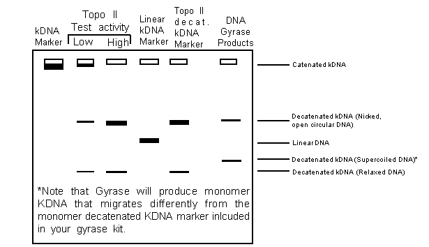
The decatenated DNA marker will be Open Circular DNA (OC, 2.5 KB monomers with at least one nick) and some Closed Circular DNA (CC) DNA. The kDNA usually contains nicked DNA monomers; thus, both OC and CC DNA will be seen. In contrast, linear DNA should not be produced since this indicates nuclease contamination. Note also that one may use the kDNA assay for DNA gyrase to identify agents that stabilize the nicked or cleavage intermediate (see link above).
Complications
The most serious complications arise when there are interfering proteins or substances in the extract being assayed. Crude, cell free extracts may contain excessive amounts of DNA binding proteins or positively charged proteins that stick to the DNA and inhibit enzyme access. Also, nuclease contaminants may degrade or nick the kDNA substrate and therefore obscure the results. A good way to deal with this problem includes cleaning up crude extracts by ammonium sulfate precipitation followed by column chromatography. Also, by diluting extracts and/or adding a tRNA carrier (to compete basic proteins), one can sometimes minimize such problems.
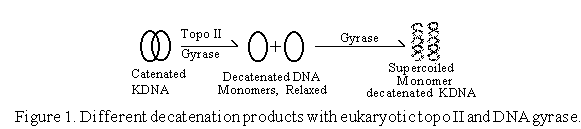
The Gyrase Assay is Based Upon a Two Step Process
1. Decatentation of kinetoplast DNA (kDNA)
2. Supercoiling of the resulting decatenated monomer kDNA species
Reaction products are resolved using a novel gel system developed by TopoGEN that allows extremely rapid and unambiguous detection of gyrase activity. The appropriate buffers, DNA substrates and DNA markers are included. Purified gyrase is not included in this kit; however, decatenated and linear DNA markers are included to allow clear and facile identification of products in the extracts or fractions defined by the user (see link above). The kDNA assay will detect poisons that stimulate DNA cleavage by gyrase as well as agents that simply inhibit catalytic activity. The advantage of a kDNA based gyrase assay is that it is fast and easy to allow activity guided purification of inhibitors from crude mixtures or extracts.
This kit may be stored at 4°C; however, for long term storage (>1 month) we recommend placing it at -20°C. Try to avoid repeated freezing and thawing of the kDNA since this will enhance degradation of the networks.