Custom Cell Based Screening Kit for NHEJ: Allows Customer to Create a Cell Line Host for an NHEJ Reporter
DNA Repair pathways in animal cells can be divided into two main categories: HR and NHEJ. HR or homologous recombination is a minor pathway but very important in protecting cells from genotoxicity. NHEJ, in contrast, is a prominent pathway in animal cells but is error prone in nature. A specific reporter based assay for NHEJ is valuable for anti-cancer drug discovery projects, learning more about the process of DNA repair and establishing intersecting pathways and druggable pathway targets.

The Custom Cell Based Screening Kit for NHEJ from TopoGEN contains reagents necessary to create a complete NHEJ reporter system in any animal cell line. It includes the NHEJ-GFP reporter with a selectable marker and a DS DNA cleavage nuclease to initiate DNA repair. Because GFP is a neutral gene reporter and not subject to selection, DNA repair can easily be examined and assayed by assessing the %GFP or GFP intensity in a population of cells. The system is mobile in that it can put into any cell lineage (or other species) to adapt to the investigators system of study. The kit optionally contains a control cell line (iHN-HeLa) that has the NHEJ-GFP reporter and a Tet-on promoter for I-Sce1. NHEJ is conveniently activated by simply adding doxycycline to the media. It serves as a control line and will help the investigator establish his own unique reporter line quickly and efficiently.
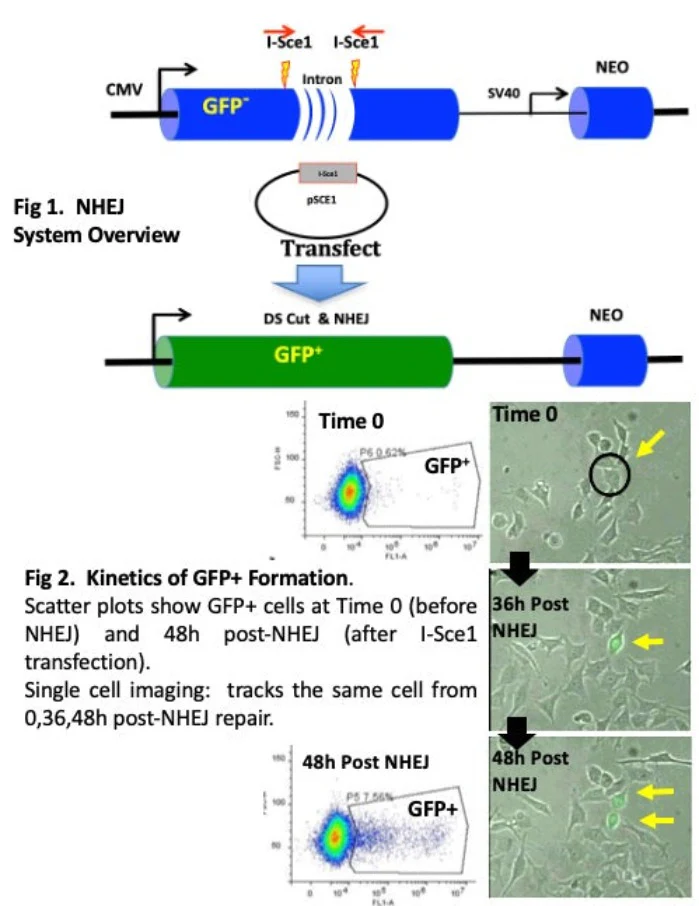
This system has been designed to allow researchers to examine and interrogate NHEJ in live cells, in real time. We designed this system to allow the customer to place the NHEJ into a cell of his or her choosing. To illustrate how the system works, we used an inducible I-Sce1 gene in a HeLa cell line (iHN-HeLa, included in the Kit). In these cells, NHEJ is conveniently activated by inducing the I-Sce1 gene under a Tet-on promoter (Fig. 1A). The GFP-NHEJ reporter contains GFP with a disrupting intron that kills the gene and renders the cells GFP negative. The intron is flanked by two I-Sce1 cleavage sites. After I-Sce1 DS DNA cleavage, incompatible end configurations are formed which are repaired by NHEJ (HR is not possible since a homologous sequence is missing). The basic assay screens for GFP+ cells that form when NHEJ faithfully restores the wild type sequence (Fig. 1B). The induction of I-Sce1 by Dox is rapid and efficient (all cells will express); however, NHEJ takes place more slowly over a period of several days, and only a small fraction of cells repair the GFP gene in an fashion that allows expression of GFP. In addition, one can use single cell imaging to follow individual GFP+ cell clones over time. This allows one to track a progenitor cell (or the 1st GFP+ cell, see Fig. 1D) and all descendants at a single cell level, if desired.